Home » Without Label » Back Muscle Diagram : Muscles Of The Back Anatomy Stock Illustration ... : Anatomynote.com found anatomy of back muscles diagram from plenty of anatomical pictures on the internet.
Back Muscle Diagram : Muscles Of The Back Anatomy Stock Illustration ... : Anatomynote.com found anatomy of back muscles diagram from plenty of anatomical pictures on the internet.
Back Muscle Diagram : Muscles Of The Back Anatomy Stock Illustration ... : Anatomynote.com found anatomy of back muscles diagram from plenty of anatomical pictures on the internet.. Some of the links in the post above are affiliate links.. We hope this picture anatomy of back muscles diagram can help you study and research. See back muscles and low back pain. As a result of overuse or strenuous activity, at times these tendons tend to get inflamed resulting in painful symptoms. Back muscles, back muscle diagram.
This can happen due to rigorous exercising, lifting heavy weights, or moving heavy items repetitively. An extremely strong tendon attached to the heel. Sore or tenderness in your lower back. The fibres attach to the clavicle, acromion and the scapula spine. Nerves in your lower back.
Muscles Diagrams: Diagram of muscles and anatomy charts ... from s-media-cache-ak0.pinimg.com Below you'll see diagrams along with the names of the back muscles that may be the cause of your pain. This can happen due to rigorous exercising, lifting heavy weights, or moving heavy items repetitively. The most common symptoms of a torn muscle, strained muscle, and pulled muscle include: It is attached to the calcaneus and is pulled by 3 flexor. The muscles of the back can be arranged into 3 categories based on their location: The back consists of the spine, spinal cord, muscles, ligaments, and nerves. Sore or tenderness in your lower back. Superficial back muscles, intermediate back muscles and intrinsic back muscles.the intrinsic muscles are named as such because their embryological development begins in the back, oppose to the superficial and intermediate back muscles which develop elsewhere and are therefore classed as extrinsic muscles.
It is the most superficial of all the back muscles.
The most common type of back pain is muscle pain—also called muscle strain or soft tissue strain. Know the causes, symptoms, treatment, recovery period of back muscle tear or injury. The muscles of the back can be arranged into 3 categories based on their location: Chart of major posterior muscles. This can happen due to rigorous exercising, lifting heavy weights, or moving heavy items repetitively. The human back extends from the buttocks to the posterior portion of the neck and shoulders. Diagram chest muscles, diagram human back muscles, diagram of back muscles and bones, diagram of back muscles and ligaments, diagram of back muscles and nerves, diagram of back muscles pain, diagram of lower back muscles, diagram shoulder muscles, human muscles, diagram chest muscles, diagram. Below you'll see diagrams along with the names of the back muscles that may be the cause of your pain. When back development is the goal, stick to one of these variations. Stiffness in the back region. Superficial, intermediate, deep and deepest layers.these muscles lie on each side of the vertebral column, deep to the thoracolumbar fascia they span the entire length of the vertebral column, extending from the cranium to the pelvis Symptoms of muscle pain include: Sore or tenderness in your lower back.
When back development is the goal, stick to one of these variations. The pelvis at the bottom of the back and the shoulders at the top of the back give the back. Creatine is now proving to be one of the most potent muscle growth accelerators giving excellent muscle mass increase and phenomenal strength increases order yours today. The deep back muscles, also called intrinsic or true back muscles, consist of four layers of muscles: Most of the time, back muscle pain is diagnosed then treated with little more than a prescription of rest, painkillers and muscle relaxants.
Labeled Anatomy Chart Of Neck And Back Muscles On White ... from media.istockphoto.com We hope this picture anatomy of back muscles diagram can help you study and research. The deep back muscles, also called intrinsic or true back muscles, consist of four layers of muscles: See back muscles and low back pain. It is attached to the calcaneus and is pulled by 3 flexor. Some of the links in the post above are affiliate links.. Diagram chest muscles, diagram human back muscles, diagram of back muscles and bones, diagram of back muscles and ligaments, diagram of back muscles and nerves, diagram of back muscles pain, diagram of lower back muscles, diagram shoulder muscles, human muscles, diagram chest muscles, diagram. Anatomy of the spine and back spine muscles diagram. While muscles like the gluteals (in the thighs) are used any time we walk or climb a step, deep back muscles and abdominal muscles are usually not actively engaged during everyday activity.
Muscle spasms (contraction or stiffening of the back muscles) muscles that feel tight;
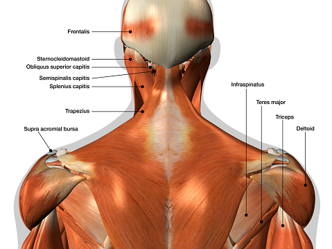
Both the deltoid and the trapezius are firmly attached to the spine of the scapula. The intermediate layer contains the erector spinae muscles, whose many functions include the extension and lateral flexion of the spine, head and neck. Muscle spasms (contraction or stiffening of the back muscles) muscles that feel tight; Deep back muscles diagram the superficial layer contains the splenius cervicis and splenius capitis muscles. They extend and rotate the head and neck. Pain increases when standing, walking, or twisting. A back muscle tear or injury similarly can be caused by overuse or strain of the muscles of the back. Superficial, intermediate, deep and deepest layers.these muscles lie on each side of the vertebral column, deep to the thoracolumbar fascia they span the entire length of the vertebral column, extending from the cranium to the pelvis See how exercise helps the back. As a result of overuse or strenuous activity, at times these tendons tend to get inflamed resulting in painful symptoms. Back muscles, back muscle diagram. The deltoid, teres major, teres minor, infraspinatus, supraspinatus (not shown) and subscapularis muscles (not shown) all extend from the scapula to the humerus and act on the shoulder joint. The trapezius and latissimus dorsi muscles connect the upper limb to the vertebral column.
Others, like sumo deadlifts, have been shown in emg studies—and in the trenches—to focus more on other muscle groups than the back. Deep back muscles diagram the superficial layer contains the splenius cervicis and splenius capitis muscles. A back muscle tear or injury similarly can be caused by overuse or strain of the muscles of the back. What is the origin and insertion of the rhomboid minor and major muscle? Pain increases when standing, walking, or twisting.
Lower Back Muscles Chart - Diagram Of Back Muscles Of The ... from i.pinimg.com Most of the time, back muscle pain is diagnosed then treated with little more than a prescription of rest, painkillers and muscle relaxants. In this image, you will find 1st cervical vertebrae, atlus, cervical plexus, 7th cervical vertebrae, 1st thoracic vertebrae, brachial plexus, spinal dura mater, filaments of spinal nerve roots, 12th thoracic vertebra, 1st lumber vertebra, iliohypogastric nerve, ilioinguinal nerve, lumbar. The muscles on the back of the trunk help lower the arms and move the body forward and sideways. Creatine is now proving to be one of the most potent muscle growth accelerators giving excellent muscle mass increase and phenomenal strength increases order yours today. It is attached to the calcaneus and is pulled by 3 flexor. Pain that radiates to your legs, buttock, or thigh areas. It is the most superficial of all the back muscles. Sore or tenderness in your lower back.
Most of the time, back muscle pain is diagnosed then treated with little more than a prescription of rest, painkillers and muscle relaxants.
Others, like sumo deadlifts, have been shown in emg studies—and in the trenches—to focus more on other muscle groups than the back. Your back hurting more when you move, less when you stay still; Muscles of the back diagram. Some of the links in the post above are affiliate links.. The muscles of the lower back help stabilize, rotate, flex, and extend the spinal column, which is a bony tower of 24 vertebrae that gives the body structure and houses the spinal cord. Chart of major posterior muscles. The pelvis at the bottom of the back and the shoulders at the top of the back give the back. Human musculature bodybuilding infographic muscular system vector human anatomy back muscle anatomy bicep male muscular anatomy human body anatomy female female anatomy muscle hamstrings muscle. Anatomy of the spine and back spine muscles diagram. We hope this picture anatomy of back muscles diagram can help you study and research. As a result of overuse or strenuous activity, at times these tendons tend to get inflamed resulting in painful symptoms. The achilles tendon in the strongest in the body. Sore or tenderness in your lower back.